Home
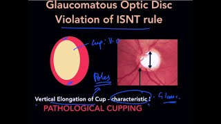
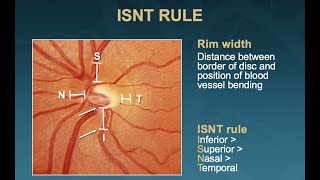
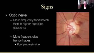
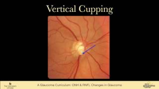
OPTIC DISC CHANGES IN GLAUCOMA
Insight Ophthalmology
29 มี.ค. 2022
การดู 35,194 ครั้ง
HOW TO READ AN OCT PRINTOUT IN GLAUCOMA || BASIC TESTING PROTOCOLS|| ( RNFL, ONH & MACULAR ANALYSIS}
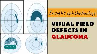
FIELD DEFECTS IN GLAUCOMA | arcuate, paracentral, nasal step, temporal wedge, baring of blindspot...
High Yield Topic : Optic Disc Changes in Glaucoma - The Complete Course
Lecture: Examining the Optic Nerve
Cup-to-disc ratio: getting it right | OT Skills Guide
iFocus Online Session #99, Glaucoma #3: Optic Disc Evaluation
The 5 Rs of Examining the Optic Disc (Malik Y. Kahook, MD)
Visual field changes in glaucoma
Lecture: General Principles of Glaucoma Management
THE BLOOD SUPPLY OF THE OPTIC NERVE | The Central retinal artery | The posterior ciliary artery
Lecture: How to Interpret OCT Findings in the Diagnostic Evaluation of Glaucoma
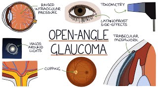
Understanding Open Angle Glaucoma
Vitreous Anatomy : All you need to know !
ANATOMY OF CORNEA made easy
9 EXAMINATION Optic Nerve Head and Nerve Fiber Layer Changes in Glaucoma
OCT in AMD( Age related macular degeneration
Macular OCT Interpretation: A Practical Discussion with Dr. David E. Lederer
Glaucoma
The Five R’s of Optic Disc Evaluation #Glaucoma #icanlearn | ECL - 35 | OOLS | Dr. Arjun Gokani
Retinal Topography | Anatomy of Peripheral Retina